Wednesday, August 24, 2016

Yale team discovers how Zika virus causes fetal brain damage

By Bill Hathaway
August 24, 2016


Human neuroepithelial stem cells self-organize into rose-like patterns and express the typical neural stem cell markers nestin (green) and SOX2 (red). They were used to understand how Zika virus infection works and to block its proliferation.

Infection by the Zika virus diverts a key protein necessary for neural cell division in the developing human fetus, thereby causing the birth defect microcephaly, a team of Yale scientists reported Aug. 24 in the journal Cell Reports.

The findings suggest that Zika virus might be susceptible to existing antiviral drugs that may prevent disruption to the developing nervous system, said the researchers.

One of the frightening side-effects of Zika virus infection in pregnant women is the risk of fetal microcephaly, in which babies are born with abnormally small brains. The multidisciplinary collaboration of Yale scientists revealed that Zika virus kills stem cells in the brain and disrupts the process of creating brain cells. An analysis shows that the virus diverts a form of the protein TBK1 from its primary job of organizing cell division to the mitochondria, the cell’s power pack, where it helps initiate an immune response. Lacking the protein at the site of cell division, cells die instead of forming new brain cells, resulting in microcephaly. The data suggest this mechanism may also contribute to microcephaly associated with other common congenital viral infections.

Researchers note that an existing FDA-approved drug, Sofosbuvir, showed promise in preventing Zika virus infection of neural stem cells in laboratory culture and also seems to keep phospho-TBK1 involved in cell division. More study needs to be conducted to prove the efficacy of the drug as a medical therapy for Zika virus, the authors said.

“There is an urgent need to identify therapeutic approaches to halt Zika infection, especially in pregnant women,” said Marco Onorati, co-first author of the paper and researcher in the lab of senior author Nenad Sestan, professor of neuroscience, comparative medicine, genetics, and psychiatry. “In the interim, we hope these findings can lead to therapies that might minimize the damage caused by this virus.”

Co-first authors of the paper are Zhen Li, Fuchen Liu, and Andre M.M. Sousa of Yale. Tamas L. Horvath and Brett Lindenbach, also of Yale, are co-senior authors of the work.

http://news.yale.edu/2016/08/24/yale-team-discovers-how-zika-virus-causes-fetal-brain-damage

Sunday, August 21, 2016

New nanotechnology “traps” viruses before they infect host cells

Tue, 01/07/2014 - 9:57am

In Figure 1, influenza viruses bind to specific carbohydrate structures on the surface of airway cells to gain entry. In Figure 2, nanotrap particles effectively mimic the cell surface so that their carbohydrate structures "trap" viruses and prevent infection.Newly emerging flu viruses could soon be countered by a treatment that Charles Stark Draper Laboratory is developing that “traps” viruses before they can infect host cells.

Further into the future, patients suffering from any type of virus could be cured with DRACO, a drug also under development at Draper that is designed to rapidly recognize and eliminate cells infected by virtually any virus.

Both methods could help safeguard against bioterrorist attacks and naturally occurring pandemics in a manner that is unlikely to lead to treatment-resistant strains. Initial testing on the treatments, which each use tiny, non-toxic particles that can be injected, inhaled, or eaten, has shown them to be effective and safe against a multitude of strains of disease.

Nanotraps, which could be taken at the first sign of infection or exposure, is likely the first of the products ready for use, and is expected to begin clinical trials in two to five years (from 01/07/2014) , according to Jim Comolli, who leads the research on the effort at Draper.

Nanotraps, developed by a team of researchers from Draper, Massachusetts Institute of Technology, the Univ. of Massachusetts Medical School, and the Univ. of Santa Barbara, are nanoparticles that act as viral “traps” using specific molecules found naturally within the human body. A paper published in the March issue of Journal of Biological Chemistry (First Published on January 28, 2013 doi: 10.1074/jbc.M112.437202) detailed their findings.

The nanotraps look like the surface of a cell, with numerous carbohydrate molecules attached that closely resemble those targeted by flu viruses in the human respiratory system. These molecules, initially characterized in the Sasisekharan Lab at MIT, act as bait for the flu virus, which bind to the nanotrap instead of a host cell and are cleared away with mucus, preventing infection, Comolli said.

The research team has demonstrated in the laboratory that the nanotraps effectively countered multiple influenza strains able to infect humans and went on to show nanotraps protected mice infected with the flu. They have also developed additional particles geared toward other types of respiratory viruses.

Nanotraps, unlike most vaccines, are not strain specific and are designed to be effective against newly emerging strains of human-adapted influenza virus. Since nanotraps mimic a fundamental step in the viral life cycle—the binding of the virus to a host cell’s receptor—nanotraps may offer an opportunity to treat devastating infectious diseases without causing the development of treatment resistance, Comolli said.

The nanotrap molecule is composed mainly of compounds found naturally in the human body so it is likely to be safe as an inhalant, topical solution, or intravenous treatment and inexpensive to manufacture. With further development, nanotraps have the potential to treat a large range of infectious diseases because the attached carbohydrates can be tuned for other viruses such as HIV, Respiratory Syncytial Virus (RSV), and Herpes Simplex Virus (HSV), as well as bacteria and toxins.

The work was originally funded by the Defense Advanced Research Projects Agency (DARPA) to address emerging pandemics or bioweapons targeting US troops; Draper is continuing the research with the intent of applying it more broadly to the civilian community.

While nanotraps could be customized to treat a variety of viruses, another project underway at Draper, DRACO could do so with a single approach. DRACO is a unique therapeutic drug that should be effective against a very broad spectrum of viruses, just as existing antibiotics are effective against a broad range of bacterial infections. DRACO could be ready for human clinical trials in five to 10 years, according to Todd Rider, who leads the research at Draper.

DRACO (Double-stranded RNA Activated Caspase Oligomerizer) is designed to detect cells that have been virally infected and then eradicate only the infected cells, rapidly ending the infection. DRACO has proven effective in vivo against influenza and three hemorrhagic fever viruses, and in vitro against 15 different viruses—including common cold viruses, the H1N1 influenza strain, adenoviruses, a mouse polio virus, dengue fever, and stomach viruses, among others. It has also been tested and proven safe in both mice and 11 different human and animal cell types representing organs like the heart, lungs, liver, and kidney, among others.

DRACO is designed to be attracted to a specific type of RNA exclusive to viral infections—long double-stranded RNA, or dsRNA. Detecting this dsRNA in a human or animal cell indicates that that host cell has been taken over by a virus and is now in the process of creating more viruses. DRACO enters cells and attaches itself to any dsRNA. Once two or more DRACOs attach to the dsRNA, they interact with one another and activate a natural self-destruct switch inside the infected cell, terminating the infected cell and the virus that it was helping to reproduce.

DRACO is designed to be both broad-spectrum and nontoxic to humans—overcoming existing issues with current anti-viral treatments. Because DRACO is so broad-spectrum and acts so rapidly, there is little opportunity for the virus to evolve defenses against this treatment, Rider said.

Rider is expanding his DRACO research and testing more strains of various viruses in cells and animals.

DRACO has been funded by grant AI057159 from the National Institute of Allergy and Infectious Diseases and the New England Regional Center of Excellence for Biodefense and Emerging Infectious Diseases, with other funding coming from DARPA, the Defense Threat Reduction Agency, and the office of the Director of Defense Research and Engineering.

Source: Charles Stark Draper Laboratory


United States Patent9,408,890
Comolli ,   et al.August 9, 2016

Multivalent liposome formulations

Abstract
This disclosure provides compositions, kits, and methods useful for treating or preventing viral and bacterial infection and reducing or preventing the effects of toxins. The methods comprise administering to a subject an effective amount of a liposomal composition.

Inventors:Comolli; James (Boxborough, MA), Trevejo; Jose (Brighton, MA), Sasisekharan; Ram (Cambridge, MA), Shriver; Zachary (Winchester, MA), Viswanathan; Karthik(Waltham, MA), Fygenson; Deborah (Santa Barbara, CA), Finberg; Robert (Newtonville, MA), Wang; Jennifer (Shrewsbury, MA)
Applicant:
NameCityStateCountryType

The Charles Stark Draper Laboratory, Inc.

Cambridge

MA

US
Assignee:The Charles Stark Draper Laboratory, Inc. (Cambridge, MA)
The Massachusetts Institute of Technology (Cambridge, MA)
University of California, Santa Barbara (UCSB) (Santa Barbara, CA)
University of Massachusetts Medical School (Boston, MA) 
Family ID:50097850
Appl. No.:14/161,555
Filed:January 22, 2014

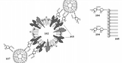
The multivalent liposome composition of the present technology has enhanced binding due to the multivalent display of binding targets and the mobility of the binding targets on the surface of the liposome. Multivalency is the display of at least two binding targets on the surface of the liposome. By way of example, but not by way of limitation, FIGS. 1A-C illustrate the enhanced inhibitory effect of multivalent liposome compositions. Referring to FIG. 1A, influenza virus 102 infects host cells by first attaching to .alpha.2-6 terminally linked SA 104 on the cell membrane 105 through HA 103. Liposomes 101 that do not contain LSTc, in general, have low effect in inhibiting influenza virus adhesion or infection. 

With reference to FIG. 1B, monovalent LSTc liposomes 106 bind to the HA receptors 103 of the influenza virus 102. 

With reference to FIG. 1C, a single multivalent LSTc liposome composition 107, e.g., with 5 mol % or more LSTc on the surface, are capable of competitively binding multiple HA receptors 203 of the influenza virus 102. The presence of multiple binding targets on the surface of a single liposome allows a single liposome to have enhanced, efficient, and high affinity binding to the virus, bacteria, or toxin as compared to the monovalent LSTc liposome of FIG. 1B. 


C.


What is claimed is: 

1. A composition comprising: a first population of lipids, a second population of lipids, cholesterol, and two or more influenza A binding targets, wherein the influenza A binding targets are linked to the first population of lipids to form BT-lipids, wherein the BT-lipids, the second population of lipids, and cholesterols form a liposome, wherein the binding targets are displayed on the outer surface of the liposome, wherein the first population of lipids and the second population of lipids in the liposome have a phase transition temperature below 41.degree. C., wherein the cholesterol comprises 15 to 30 mol % of the liposome. 


Patent Family:


Inventing Virus Traps

Viruses will bind to any cell possessing the correct receptor. This leaves them vulnerable to engineered cells expressing the correct receptor, but are otherwise not viable for productive infection. In work published in Ecology Letters, we described a mathematical model to predict the effectiveness of virus traps in driving viruses to extinction. We validated the model using bacteriophage Φ6 and a naturally occurring trap host. We found that traps could indeed drive viruses to extinction. The figure below accompanied a New York Times article about the work.

viral-lg
United States Patent Application20060018912
Kind CodeA1
Finberg; Robert W. ;   et al.January 26, 2006

Snares for pathogenic or infectious agents and uses related thereto 
Abstract
The present invention provides a snare molecules comprising an attachment moiety (which facilitates attachment of a receptor to a cell) and a receptor for a toxic pathogenic or infectious agent, e.g., a virus. Methods of producing such snare molecules and their therapeutic and/or prophylactic uses are also provided by the present invention

Inventors:Finberg; Robert W.(Sudbury, MA) ; Asher; Damon R.(Holden, MA)
Correspondence Address:
    LAHIVE & COCKFIELD, LLP.
    28 STATE STREET
    BOSTON
    MA
    02109
    US
Assignee:UNIVERSITY OF MASSACHUSETTS
Boston
MA
Family ID:35285443
Appl. No.:11/139272
Filed:May 27, 2005

Related U.S. Patent Documents

Application NumberFiling DatePatent Number
60575149May 28, 2004
60661991Mar 11, 2005

Current U.S. Class:424/178.1 ; 530/391.1
Current CPC Class:C07K 16/1009 20130101; C07K 14/705 20130101
Class at Publication:424/178.1 ; 530/391.1
International Class:A61K 39/395 20060101 A61K039/395; C07K 16/46 20060101 C07K016/46

Claims

1. A snare molecule, comprising an attachment moiety that facilitates attachment of the molecule to a blood cell and a receptor for a pathogenic agent, wherein the receptor is not naturally found on the blood cell. 
From PAIR:
07-05-2016NOANotice of Allowance and Fees Due (PTOL-85)

I expect the US patent to issue shortly!

Friday, August 19, 2016

BRAZIL HEALTH OFFICIALS: ZIKA VIRUS IS NOT RESPONSIBLE FOR RISE IN BIRTH DEFECTS, IT’S SOMETHING ELSE


ALEXA ERICKSON
AUGUST 18, 2016

Fear surrounding the threat of Zika virus has plagued the media, and its steady spread across Latin America and Pacific island nations — along with its inevitable arrival in parts of the continental U.S. — has many worried. The mysterious virus, suspected of causing birth defects, has provoked the U.S. Centers for Disease Control and Prevention to update its recommendations on pregnancy and sex. But there are plenty of skeptics wondering what we should and shouldn’t believe. Now, there seems to be a breakthrough.

Amidst the growing concerns, health officials in Brazil have admitted that Zika alone may not be responsible for the rise in birth defects in parts of the country.

The virus may be linked to the birth defect called microcephaly, but while Zika has been spreading at extremely high rates throughout Brazil, microcephaly has not.

“We suspect that something more than Zika virus is causing the high intensity and severity of cases,”explains Dr. Fatima Marinho, Director of Information and Health Analysis at Brazil’s Ministry of Health.

Here are some facts you should know:

Almost All Cases Of Brazil Microcephaly Took Place In The Northeast


Since last November, Brazil has seen more than 1,700 cases of microcephaly or other birth defects of the central nervous system. When the cases were first detected, health officials believed they’d witness “an explosion of birth defects” across the country, but that wasn’t the case. In fact, data compiled by Marinho and colleagues found that socio-economic factors may be involved, since the majority of the women who gave birth to babies with microcephaly were poor and resided in small cities or on the outskirts of big cities. The outbreak also occurred in extremely poverty-stricken areas of Brazil that use massive amount of banned pesticides. Environmental pollution and toxic pesticide exposure have been linked to many adverse health effects, including birth defects

There Is Not Enough Data

Not enough data can rightfully link the Zika virus to microcephaly, and the data that does exist mainly comes from incomplete hospital reports, with tests to confirm the virus often not carried out.

It’s even been suggested that microcephaly may be the combination of Zika along with other infections such as dengue and chikungunya, with the Brazilian doctor who first reportedly established the link between Zika virus and microcephaly now claiming Bovine viral diarrhea virus (BVDV) may be involved.

Colombian Women and Zika Virus


The New England Complex Systems Institute (NECSI) followed nearly 12,000 pregnant Colombian women infected with Zika virus and found no cases of microcephaly, yet four cases of microcephaly were reported among women who had Zika infection with no symptoms and were therefore not included in the study.

According to NECSI:

This gives a consistent interpretation that there is no direct link between Zika and microcephaly except for random co-occurrence. We note that the base rate of microcephaly in the absence of Zika is 140 per year in Colombia, which is consistent with the approximately 50 microcephaly cases in the first 4 months of 2016, only 4 of which have been connected to Zika. When interpreting Zika as the cause, background cases must be subtracted.


WHO Expert Weighs In

According to Florence Fouque, a World Health Organization (WHO) expert on animals that carry viruses, the public response to the Zika virus is “completely hysterical.” She said the hysteria comes from the findings of the virus harming pregnant women, and that it can be sexually transmitted.

“It’s like AIDS,” she said. “People make this link and that’s why they are really afraid.

Oliver Brady, an epidemiologist with the London School of Hygiene and Tropical Medicine who was asked by Brazilian officials to assess the Zika-microcephaly situation, also weighed in on the subject:

You see that with a lot of arboviruses [viruses spread by mosquitoes and other insects]. . . .They have pathogenic qualities and if you put them in the right tissue then they will cause some sort of damage. And they tend to be quite transmissible across a variety of barriers anyway. So it doesn’t necessarily mean that that’s the mechanism that’s happening out there in the field, even if it does work in the lab.

The U.S. Is Ignoring Valuable Data

Among those jumping to conclusions is the U.S., which, despite the data, has rushed to launch a clinical trial of an experimental Zika vaccine without conclusive proof that Zika causes microcephaly.

The trial will involve 80 healthy volunteers between the ages of 18 and 35. These participants will be vaccinated with varying doses of the experimental vaccine, and placebos will be given.

There are concerns about such a vaccine, Dr. Scott B. Halstead, former senior adviser of the Dengue Vaccine Initiative and the founder of Children’s Vaccine Initiative, told the University of Minnesota Center for Infectious Disease Research and Policy:

It’s happened. We have a vaccine that enhances dengue. . . . It’s clear as the nose on my face: Vaccine recipients less than 5 years old had five to seven times more rates of hospitalizations for severe dengue virus than placebo controls. Halstead is specifically referring to a three-year study that suggested the vaccine causes antibody-dependent enhancement (ADE). He said: “Over time, you make and keep protective levels of antibody from the initial infection, but you lose the cross-reactive antibodies. . . . That allows a second dengue infection to cause severe illness.”

The University of Minnesota Center for Infectious Disease Research and Policy sided with concerns discussed by Dr. Philip K. Russell, the former director of the Walter Reed Army Institute of Research and commander of the U.S. Army Medical Research and Development Command, as well as founding president and chairman of the Sabin Vaccine Institute: “Russell said that the fact that Zika is occurring in areas where dengue has been endemic hints at a serious potential problem with ADE and Zika vaccine development.” Russell himself noted, “The current epidemic of Zika, which is usually mild disease, is made a lot worse in these populations,” and “I think there’s a major effect, but the studies haven’t been done yet to sort that out.”

Toxic Exposures For Battling Zika

Due to the media frenzy, many areas have increased their pesticide use as a means for combating the Zika virus. And while a Clean Water Act permit is typically required to spray pesticides in areas where they might end up in water, the Zika virus has been used as an excuse to further harm the planet and its inhabitants.

The Zika Vector Control Act was passed by the House of Representatives, exempting pesticide applicators from needing a Clean Water Act permit, even when spraying near water. But opponents say the bill has nothing to do with battling Zika, claiming it has been on the table for years, with the majority fighting to slap any name on it to make it convenient to the time.

Research has shown the detriments to such spraying, like the evidence presented at the Pediatric Academic Societies 2016 Meeting, which explained that aerial pesticide exposure is linked to an increased risk of developmental delays and autism spectrum disorder among children

Mosquito Experts Know The Truth

Even mosquito experts suggest things aren’t right. According to Chris Barker, Ph.D., a researcher of mosquito-borne viruses at the University of California, Davis School of Veterinary Medicine: “I think the risk for Zika actually setting up transmission cycles that become established in the continental U.S. is near zero.” He anticipates the virus to mimic other tropical diseases spread by mosquitoes, like dengue fever and chikungunya, by making its way into the U.S. with small clusters of outbreaks in Southern states and not much activity elsewhere.

http://www.collective-evolution.com/2016/08/18/brazil-health-officials-suspect-zika-virus-is-not-responsible-for-the-rise-in-birth-defects/

A pregnant woman's immune response could lead to brain disorders in her kids

August 19, 2016 by Myka Estes, The Conversation


Human brain cells. Spike Walker, Wellcome Images, CC BY-NC-ND

Pregnant women, like everyone, get sick. And like everyone else, their bodies try to fight infection and, importantly, keep it from reaching the growing fetus.

If the mother's immune system successfully defeats the virus before the developing baby is exposed or if the virus never crosses the placenta, is harm averted?

Counterintuitively, this protective response may be a risk factor for some neurological conditions in the baby later on.

This is the question that researchers, including me, have been probing at the neurodevelopment lab at the University of California, Davis. Research suggests that the mother's defensive immune response to an infection, for instance, alone is sufficient to cause lifelong changes in brain architecture and function and in behavior in the offspring. This response is a strong risk factor for brain disorders like autism and schizophrenia.

Kimberley McAllister, who leads the neurodevelopment lab, and I reviewed recent research about what is called maternal immune activation (MIA) in humans and in animals in an article published today in Science. So what do we know so far about MIA and where research is headed?

What is maternal immune activation?

Maternal immune activation refers to the mother's immune system defensive response to invading pathogens. During pregnancy the immune system changes to accommodate the needs of the growing fetus. These changes are complex and depend on her stage of pregnancy and the pathogens she encounters. The intensity of the immune response is highly individualized and represents a complex interaction between the mother's genes and environment.

A study in humans has suggested that the degree and duration of MIA determines the risk of a child being diagnosed with a brain disorder later on, like autism or schizophrenia. And infections aren't the only cause of MIA. For example, an increased risk is also associated with psychological stress during pregnancy, which triggers a similar immune activation.

Studies like these identify associations, but not causation. However, animal studies support a causal role for these risk factors and are beginning to reveal the underlying mechanisms.

Why does MIA increase the risk of brain disorders?

Every pathogen has its own unique signature and trick up its sleeve to subvert the body's defenses. However, at least during the first days of infection, the immune system initiates a standard response. We are all familiar with its consequences: fever, lethargy, aches and pains.

While the body weathers these symptoms, the immune system is hard at work deploying communicative proteins called cytokines, which tell immune cells where to go and how to destroy invading pathogens.

During an infection, the concentrations of cytokines change rapidly in order to guide the immune response. Too little and you succumb to the pathogen; too much and youdie of sepsis. And besides telling immune cells where to go and what to do, cytokines have also been shown to play integral roles in brain development in animal models.

During pregnancy, the mother's cytokines can affect the fetus.

In the developing brain, the concentration of cytokines is tightly regulated. Here, too little or too much, in the wrong place or at the wrong time, can alter the architecture and function of the developing brain.

While the full repertoire of these changes are unknown, studies using animals have shown that altering cytokine concentrations changes how brain regions are connected to one another and how they communicate. These types of alterations in specific areas of the brain are thought to underlie numerous brain disorders.

Our lab has shown that these cytokine changes in the fetal brain are lifelong and region-specific. Another recent paper showed that a greater concentration of a specific cytokine, IL-17, within the fetal brains of mice was sufficient to lead to brain changes, which resulted in altered behaviors associated with autism and schizophrenia. Cytokine changes are also associated with another indicator of brain disorders:alterations in the number of connections made between neurons.

Whether or not these cytokine changes are a cause of these disorders in humans remains to be seen. But there is evidence from post-mortem brain samples taken from individuals with autism and schizophrenia that show altered concentrations of cytokines.

The ultimate goal is to identify a cytokine profile that predicts certain behaviors and neurological disorders in animals. In the future, we could potentially use this profile to identify biomarkers of specific brain disorders in people.

MIA, genetics and environment

Over the past decade, hundreds of genes have been associated with a wide range of brain disorders. However, each of these genes increases risk only slightly. Any number of combinations of the associated genes may result in a brain disorder such as autism.

To us, research suggests that the maternal infection acts as a disease primer. In other words, maternal immune activation may make the fetus more susceptible to genetic and environmental risk factors.

Hypothetically, if the fetus experiences a low dose of MIA, and as an adolescent experiences stress or uses cannabis, the combination might trigger brain changes that could lead to schizophrenia.

Researchers are just starting to explore how genetic and environmental risk factors combine with MIA. These studies will help to explain why the outcomes of MIA are so diverse. The good news is that the majority of cases do not lead to any observable disorders.

While this may sound like a daunting challenge, important progress is being made. For example, many of the genes we know to be associated with certain conditions appear to be related to one another in how they function in the brain, which gives us insight into what brain processes are disrupted when people have these disorders.

This may explain the role cytokines play in raising risk for these disorders. They transmit messages from one cell or tissue to another, triggering numerous changes within the receiving cell. The processes and pathways in the brain that cytokines may disrupt may be the same processes and pathways that the risk genes disrupt as well.

If this is the case, it suggests we may be able to develop new therapeutics that benefit individuals with different disorders, regardless of whether the cause of the disorder is primarily environmental, like a maternal infection, or genetic.

Where is this research headed?

Numerous questions remain unanswered in our field. Why do only some children born to women who experience MIA go on to develop these disorders? How can we identify high-risk pregnancies? What combination of genetics and environmental risk factors distinguish autism from schizophrenia or Alzheimer's?

All of these questions are currently being explored using animals, and the initial findings are both astounding and hopeful. For example, some behavioral features of these disorders can be prevented with early intervention or even reversed autism-like disorders in adulthood.

Moreover, some of these interventions are noninvasive and involve altering gutmicrobiota and immune system signaling. Whether or not similar approaches will work in humans remains to be seen.

If MIA does indeed act as a disease primer for a wide range of brain disorders, it is imperative that we identify groups at higher risk and develop therapeutic interventions. Climate change, population growth and urbanization all increase the risk of exposure to pathogens. The social, economic and emotional tolls are too substantial to ignore.

MIA risk has always been there. It is only our understanding that is new. By studying MIA, we may now begin to reveal common principles underlying seemingly disparatebrain disorders. And most hopefully, we may harness these insights to increase our resilience.

Explore further: Immune activation in pregnant mice affects offspring, potential implications for neurodevelopmental disorders

Provided by: The Conversation

http://medicalxpress.com/news/2016-08-pregnant-woman-immune-response-brain.html

How gastro-causing virus latches onto and infects cells

It's the most common viral cause of diarrhoea worldwide, but norovirus is notoriously tough to study. Scientists in the US used a mouse version of the disease to uncover how it gets into cells. Belinda Smith reports.


The dreaded gastro: it sweeps through workplaces, schools, restaurants, resorts – anywhere with loads of people. And while cramps, diarrhoea, vomiting and a general feeling of malaise usually last just a couple of days, how the bug infects people and causes disease is still largely a mystery.
Now, biologists in the US have found a clue. They uncovered the pathogen’s gateway in mice – a protein hanging off the outside of cells to which the virus can latch and sneak inside to replicate. Without the protein, called CD300lf, the virus is unable to invade the cells.
Around 20 million people in the US are struck by acute gastroenteritis each year, leading to 400,000 emergency department visits and hundreds of deaths – mainly among young children and the elderly – all thanks to norovirus, a tiny virus behind 90% of non-bacterial gastroenteritis.
The disease can be severe, or even fatal, in patients already hospitalised or in a nursing home. This isn't helped by the fact that the illness is extremely contagious – even swallowing virus particles in the air after an infected person has vomited is enough to bring someone down with the disease.
But unravelling the cellular mechanisms behind the disease’s virulence has been hampered by the fact that norovirus is really hard to grow in the lab.
And strains of norovirus are particular to their host species. Scientists can’t infect a mouse or rat with human norovirus. (If you find yourself struck down with norovirus, for instance, it won’t spread to your pets – just other people with whom you come in contact.)While it’ll happily infect and spread through people in real life, it turns its nose up at human cells in a petri dish.
So when, in 2003, a mouse version of norovirus was discovered by scientists at the Washington University in the US, biologists finally had an animal model to study the disease.
Under the microscope, mouse and human norovirus look similar – spherical protein shell around 30 nanometres wide encasing genetic material.
Herbert Virgin, part of the team that found the mouse norovirus as well as the current study, along with Robert Orchard and Craig Wilen, also the Washington University, wanted to find out why the mouse norovirus targeted only mice – and this, perhaps, could point to how the human version infects us.
Using a gene-editing technique called CRISPR-Cas9, they looked for mouse genes that enable norovirus infection by snipping them out and seeing what happened.
They found mouse cells lacking the gene CD300lf were immune to the disease. CD300lf codes for CD300lf, a protein that sits on the surface of mouse cells and Orchard, Wilen and their colleagues believed it key to norovirus infection.
When they inserted the CD300lf gene into the DNA of human cells in a dish, which then began expressing the CD300lf protein, the mouse norovirus had no issue infecting and multiplying inside them too.
"This tells us that the species restriction is due to the ability to get inside the cells in the first place,” Virgin says.
“Once inside the cells, most likely all the other mechanisms are conserved between human and mouse noroviruses since the viruses are so similar."
Strangely, the norovirus needed another molecule – or “co-factor” – to infect cells, but the team was unable to establish its identity.
Orchard thinks it’s probably not a protein, but a small molecule found in blood.
This mystery molecule may be crucial to cajoling the virus to grow in human cells in the lab – and one day, let scientists develop drugs that disrupt the disease's infection route.

Humans caught the cold from CAMELS: Virus came from the same animals that passed us the deadly MERS

  • First human to be infected with the common cold caught it from a camel 
  • This is the same way Middle East Respiratory Syndrome was passed over
  • Researchers warn the way the cold spread could be repeated with MERS
  • This could lead to a global outbreak of the deadly respiratory condition 
The virus that causes the common cold was originally passed to humans from a camel, according to a new study. 
This means the virus came from the desert-dwelling animal that also passed us the deadly Middle East Respiratory Syndrome (MERS), and the researchers warn MERS might spread across the globe in the same way the common cold has.

The virus that causes the common cold was originally passed to humans from a camel, according to a new study. This means the virus came from the desert-dwelling animal that also passed us the deadly Middle East Respiratory Syndrome (MERS)

CAMELS GAVE US THE COLD 

The virus that causes the common cold was originally passed to humans from a camel.
Researchers had been looking into the origins of the MERS virus when they made the discovery.
The scientists took samples of the camels' cold viruses and discovered they were also capable of infecting humans.
Further analysis of viruses in bats, humans and dromedaries showed the common cold virus was transmitted from camels to humans.
Researchers at the University Hospital of Bonn in Germany had been looking into the origins of MERS when they made the discovery.
'In our MERS investigations we examined about 1,000 camels for coronaviruses and were surprised to find pathogens that are related to HCoV-229E, the human common cold virus, in almost six per cent of the cases,' said Professor Christian Drosten lead author of the research.
The scientists took samples of the camels' cold viruses and discovered they were also capable of infecting humans.
The researchers reported their findings in a paper published in the Proceedings of the National Academy of Sciences. 
The scientists took samples of the camels' cold viruses and discovered they were also capable of infecting humans. Coronaviruses are a group of viruses responsible for causing the common cold, pictured
Further analysis of viruses in bats, humans and dromedaries showed the common cold virus was transmitted from camels to humans.
They found the human immune system is able to defend itself against the camel viruses, just as it can against common cold viruses - meaning there is no threat of an epidemic.
But the future is less certain for MERS.
'The MERS virus is a strange pathogen: smaller, regionally restricted outbreaks, for example in hospitals, keep occurring,' said Professor Drosten.
'Fortunately, the virus has not adapted well enough to humans, and has consequently been unable to spread globally up to now.'
The global spread of HCoV-229E happened through human-to-human transmission, which is highly likely to have occurred during a past pandemic.
This means there is a concern the same could happen with MERS. 
'Our current study gives us a warning sign regarding the risk of a MERS pandemic—because MERS could perhaps do what HCoV-229E did,' Professor Drosten added.
There is need for action, the researchers said. 
A team from the same hospital are working intensively on researching a vaccine against MERS which will go into clinical testing early next year.

WHAT IS THE MERS VIRUS?

MERS is a viral respiratory disease caused by a coronavirus (MERS-CoV).
It was first identified in Saudi Arabia in 2012. 
The virus can affect humans as well as animals though scientists do not yet know exactly how people become infected with MERS-CoV.
It is a type of coronavirus - a large family of viruses that can cause diseases ranging from the common cold to Severe Acute Respiratory Syndrome (SARS).
Typically a person infected with MERS will suffer a fever, cough and/pr shortness of breath.
Pneumonia is a common sign, as well as gastrointestinal symptoms, including diarrhoea.
In severe cases the virus can cause respiratory failure that requires ventilation and support in an intensive care unit.
The virus is known to cause more severe disease in those people who already have a weakened immune system, older people, and those with chronic disease such as diabetes, cancer and chronic lung disease.  
It is a zoonotic virus, and it is thought humans can become infected via direct or indirect contact with camels that have the disease. 
Human-to-human transmission is possible, but only via very close contact.
Source: WHO
MERS is a viral respiratory disease caused by a coronavirus (MERS-CoV, oictured). It was first identified in Saudi Arabia in 2012. The virus can affect humans as well as animals though scientists do not yet know exactly how people become infected with MERS-CoV

Zika virus harms adult brain cells in mice

First study on the disease's effects on adult brain cells suggests risk may not be limited to foetuses. Amy Middleton reports.

The Zika virus has spread through Central and South America, and is becoming more prevalent across the globe – largely thanks to mosquitos.

The Zika virus might not just harm the brain of the developing foetus – it could also affect parts of the adult brain, a new study in mice suggests.
A published in Cell Stem Cell by US researchers found the virus targets stem cells in parts of a fully grown mouse brain responsible for learning and memory and stops them proliferating. The team suggests the virus should be monitored in every patient – not just pregnant women.
"Based on our findings, getting infected with Zika as an adult may not be as innocuous as people think," says Joseph Gleeson, a neurologist at Rockefeller University in New York and an author of the paper.
The Zika virus is a mosquito-borne and sexually transmitted virus first reported in the 1950s. Back then, it was contained within a narrow area along the equator, but the virus spread in 2007 to the Americas, eventually creating an epidemic the World Health Organisation has called a “Public Health Emergency of International Concern”.

Most recent attention in Zika research to date has centred on pregnant women. This is because the virus can spread to foetuses, causing severe brain defects including microcephaly – a condition that causes babies to be born with an unusually small head.
Now, a new study by researchers at Rockefeller University and La Jolla Institute in San Diego suggests the virus might also put adult brains at serious risk.
Microcephaly is the result of interruption during a very important process in a foetus’ brain development: when stem cells divide to turn into fully functioning neurons.
Some of these pre-neuron stem cells stick around in the adult brain, too. In mice, they exist in two regions that are important for learning and memory: the subventricular zone of the anterior forebrain and the subgranular zone of the hippocampus.
Interruptions to the neuron development process in adult stem cells have been linked to Alzheimer’s disease and cognitive deficits similar to the symptoms of depression.
While it’s not known exactly how Zika leads to microcephaly, the research team hypothesised that this progression could also potentially play out in stem cells in the adult brain.
To test this theory, the team monitored the brains of mice infected with Zika, using glowing biomarkers to track the virus and measure affected areas of the brain.


The fluorescent biomarker (in green) revealed that the adult mouse brain could be infected by Zika in a region called the subgranular zone of the hippocampus.
LABORATORY OF PEDIATRIC BRAIN DISEASE AT THE ROCKEFELLER UNIVERSITY / CELL STEM CELL
According to their results, which Gleeson says were “dramatic”, the virus targeted the two areas containing stem cells and stopped the cells dividing.
"In the parts of the brain that lit up, it was like a Christmas tree," Gleeson explains.
"It was very clear that the virus wasn't affecting the whole brain evenly, like people are seeing in the foetus. In the adult, it's only these two populations that are very specific to the stem cells that are affected by virus. These cells are special, and somehow very susceptible to the infection."

The long-term effects of Zika on adult brains are still unknown, and the team says more research is needed to ascertain whether the mouse model echoes the virus’ effect on humans. Nevertheless, the researchers consider their results a warning sign.

"The virus seems to be travelling quite a bit as people move around the world," says Gleeson.

"Given this study, I think the public health enterprise should consider monitoring for Zika infections in all groups, not just pregnant women."